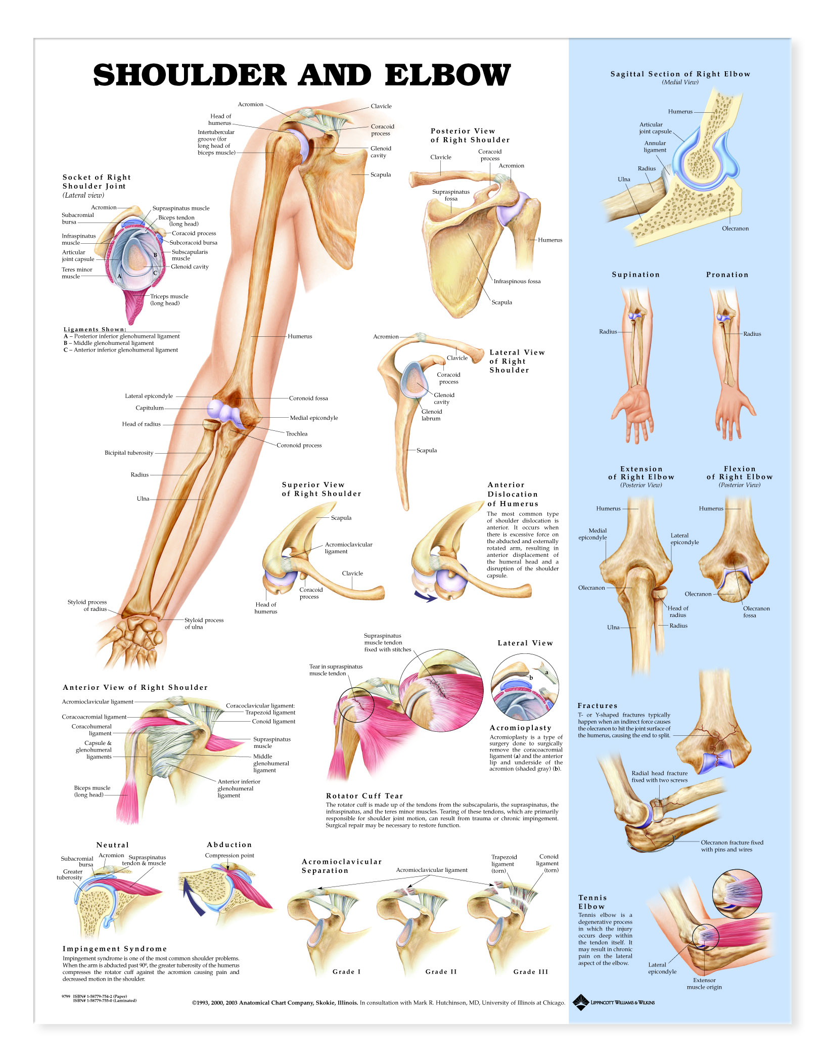
Illustrates normal shoulder and elbow anatomy.
Detailed labeled illustrations of the shoulder as follows:
general bone structure and anatomy of the shoulder and elbow
detailed view of the socket of the right shoulder joint
posterior, lateral, and superior views of the bones of the right shoulder
anterior view of the right shoulder with ligaments, bones and major muscles
Also illustrates and describes some common injuries of the shoulder:
anterior dislocation of humerus
rotator cuff tear
impingement syndrome
acromioclavicular separation
Illustrations of the elbow include:
sagittal section (medial view) of the right elbow
supination and pronation
superior views of extension and flexion of the right elbow
Also illustrates and describes common fractures of the elbow and tennis elbow
Made in the USA.
UNMOUNTED - Printed on high-quality, heavy paper. Get an unmounted chart if you are going to frame it, or if you just want to tape or tack it up on a wall. Our human anatomical charts measure 20W x 26H. (Note: Unmounted charts cannot be displayed on the chart stand).