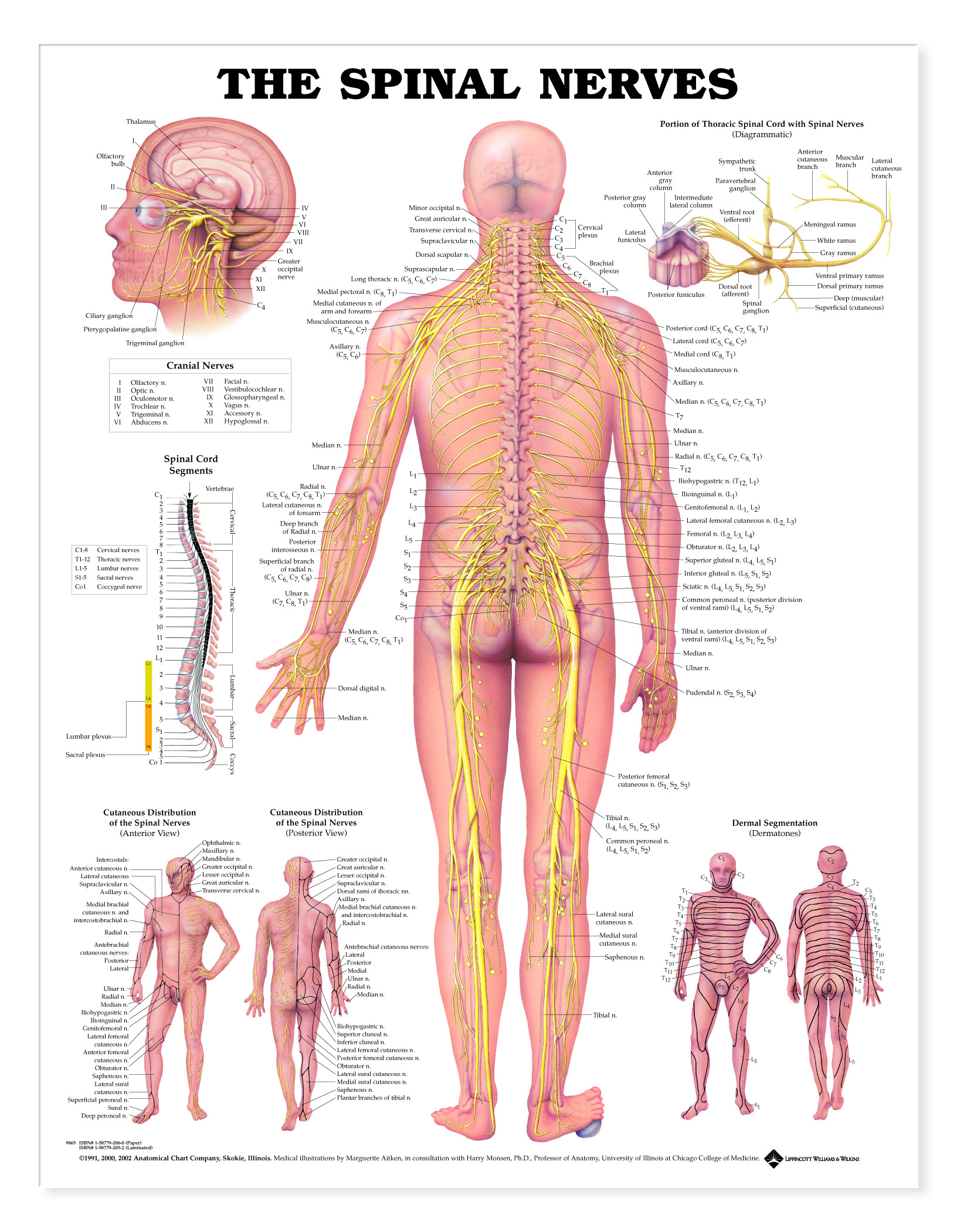
Illustrates the spinal nerves and pathways through the body.
Central illustration shows a posterior view of the spinal nerves exiting from the vertebral column and running throughout the body. Important skeletal structures are included. All nerves and their corresponding vertebra are clearly labeled. Also includes detailed illustration of the cranial nerves; diagrams the portion of the thoracic spinal cord with spinal nerves, spinal cord segments, anterior and posterior cutaneous distribution of spinal nerves and dermal segmentation (dermatomes).
The stone under the foot illustrates the changes in the nerves of a flexed foot.
Made in the USA.
FLEXIBLE LAMINATION - Flexible plastic 1.5 mil lamination with metal eyelets in each top corner for convenient wall hanging or for use with chart stand; markable (write-on/wipe-off). If you are going to put the chart on an office or classroom wall, this will hang nicely without curling at the edges. Also, the chart will easily roll up, if you want to carry it with you. Our most popular mounting. Our human anatomical charts measure 20W x 26H.