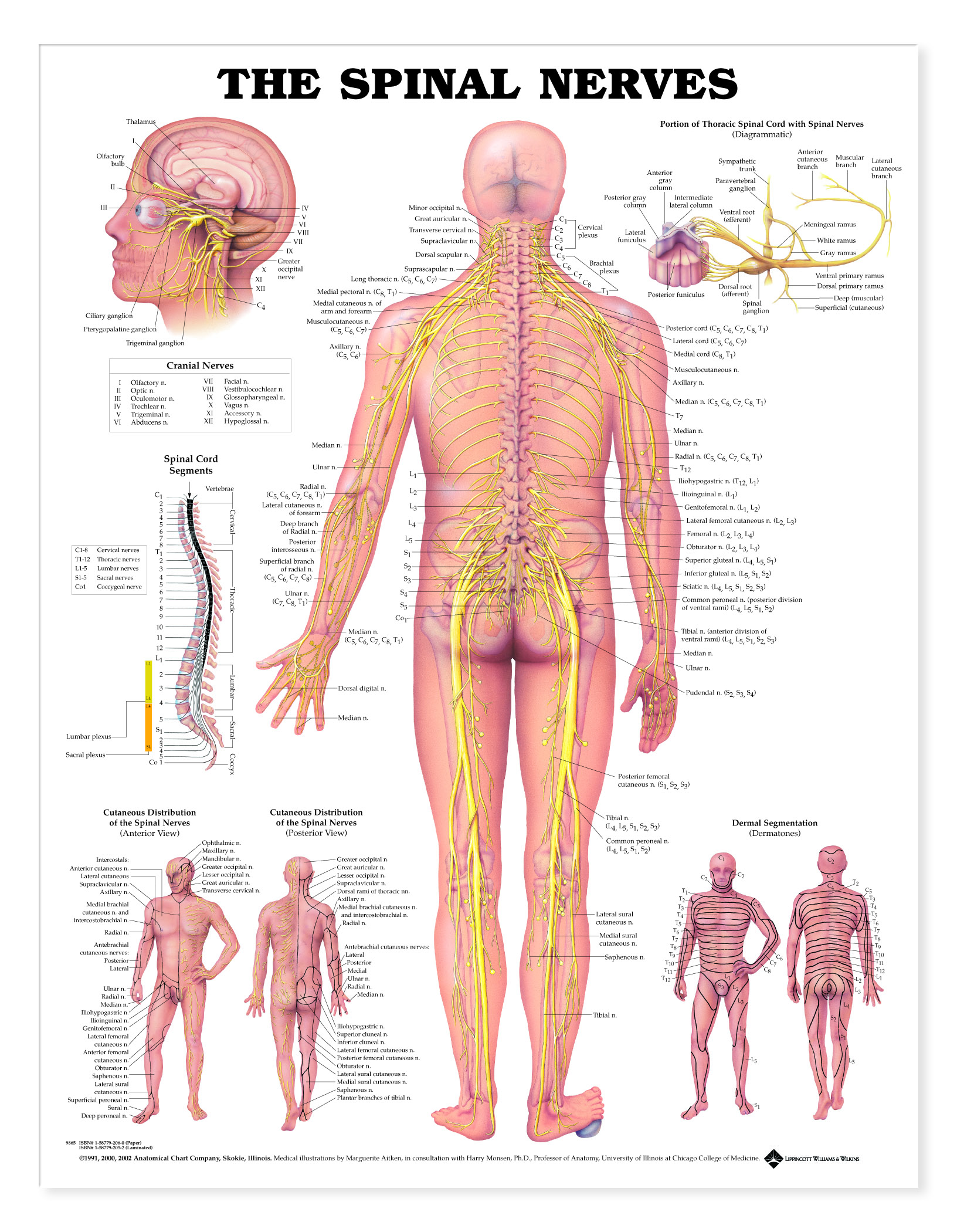
Illustrates the spinal nerves and pathways through the body.
Central illustration shows a posterior view of the spinal nerves exiting from the vertebral column and running throughout the body. Important skeletal structures are included. All nerves and their corresponding vertebra are clearly labeled. Also includes detailed illustration of the cranial nerves; diagrams the portion of the thoracic spinal cord with spinal nerves, spinal cord segments, anterior and posterior cutaneous distribution of spinal nerves and dermal segmentation (dermatomes).
The stone under the foot illustrates the changes in the nerves of a flexed foot.
Made in the USA.
UNMOUNTED - Printed on high-quality, heavy paper. Get an unmounted chart if you are going to frame it, or if you just want to tape or tack it up on a wall. Our human anatomical charts measure 20W x 26H. (Note: Unmounted charts cannot be displayed on the chart stand).